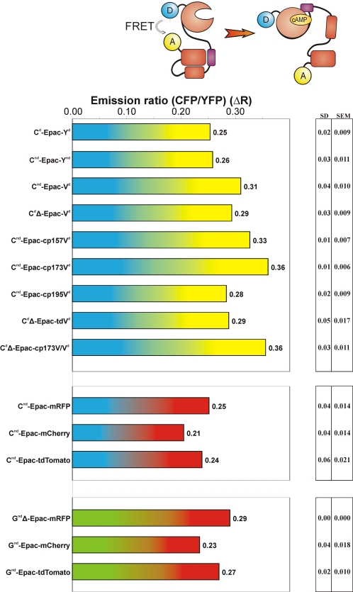
Figure 3. FRET span in cAMP sensors.
The indicated constructs were expressed in HEK-293 cells and assayed for cAMP-induced changes in donor to acceptor ratio on a fluorescence microscope equipped with dual photometers. Donor and acceptor emission were read out simultaneously, and the baseline ratio was set to 1.0 at the onset. FRET span ΔR was determined by calculating the ratio change following addition of IBMX and Forskolin. This raises intracellular cAMP levels maximally and saturates the sensor. For further detail, see the text and Methods.

