
Figure 2. Histology of hippocampal sections before and after laser microdissection.
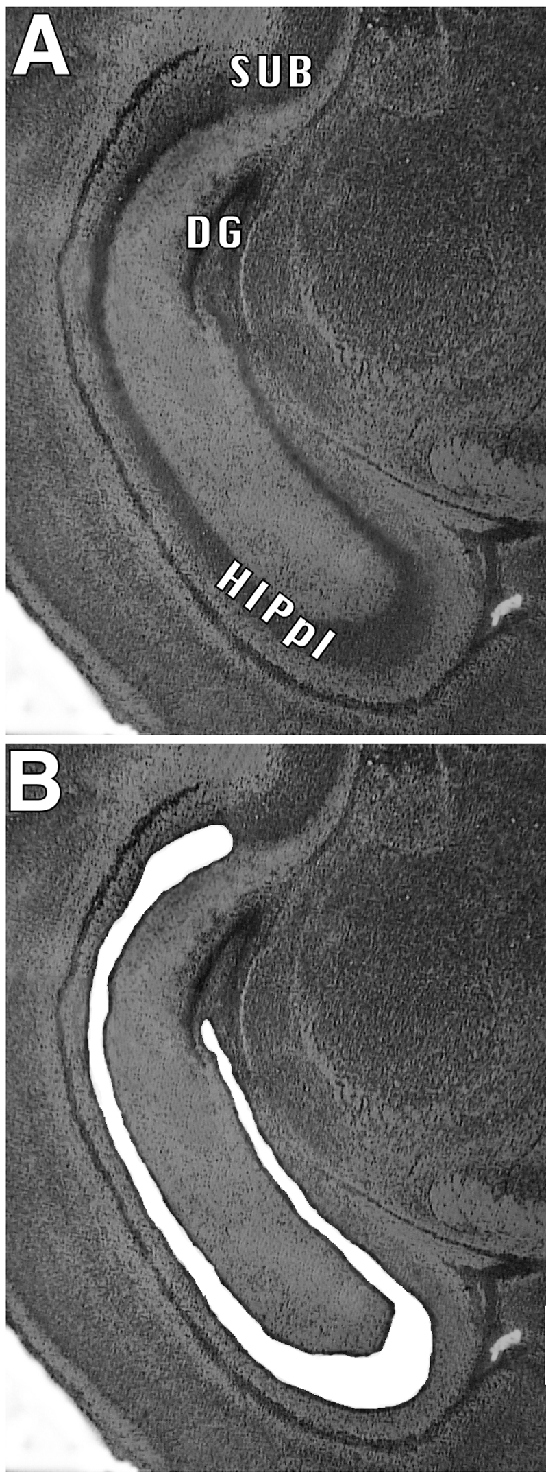
Photomicrograph of a typical HistoGene-stained coronal section through the hippocampus of a 3-day-old mouse (a), and the same section following excision of the hippocampal pyramidal layer by microdissection on a Leica ASLMD laser capture microdissection system (b). HIPpl, hippocampal pyramidal layer; DG, dentate gyrus; SUB, subiculum. Note the precision of the cut.