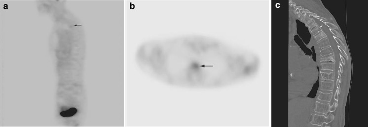
Fig. 4.
a Benign subacute compression fractures in a patient without history of malignancy. Sagittal FDG-PET image demonstrates mildly increased radiotracer uptake in the upper thoracic spine (SUV = 1.8; arrow). b Axial FDG-PET image at the level of T4 demonstrates mildly increased radiotracer uptake (arrow), consistent with subacute compression fracture. c Sagittal CT image demonstrates compression fracture of T4 (arrow) and multiple chronic osteoporotic compression fractures throughout the thoracic spine

