
FIGURE 2. Selective hydrolysis of HGF by mast cell and neutrophil peptidases.
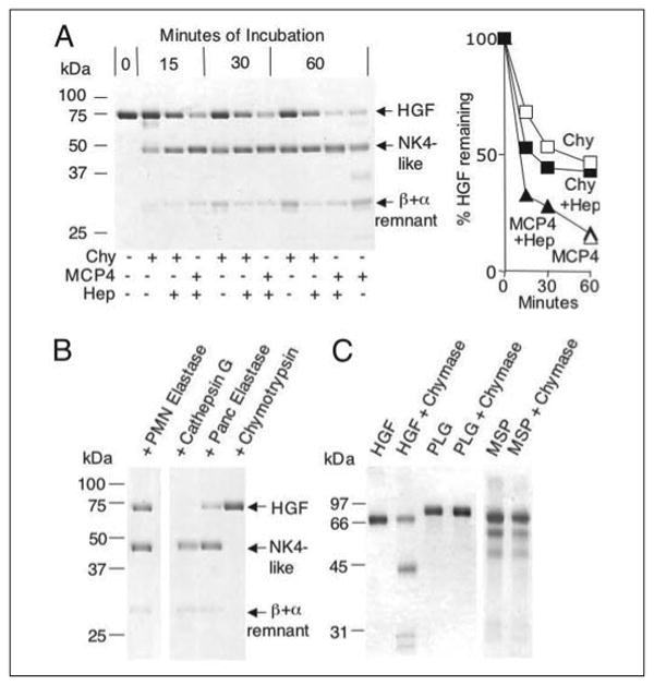
A, shows non-reducing SDS-PAGE of products generated from human HGF by the chymotryptic peptidases human chymase (Chy) and mouse MCP4, with time-dependent generation of an NK4-like major fragment and a β chain attached to an α remnant. Incubation intervals are indicated. Some incubation mixtures contain heparin (Hep). Incubation of HGF alone for 60 min does not yield visible fragmentation (not shown). The graph shows results of densitometry. B, shows results of incubation with neutrophil (PMN) elastase, cathepsin G, pancreatic elastase, and chymotrypsin, revealing that PMN elastase and cathepsin G generate an NK4-like fragment similar to classical NK4 generated by pancreatic elastase. Chymotrypsin has comparatively little activity. C, shows results of incubating chymase with HGF-related proteins, including MSP and plasminogen (PLG).