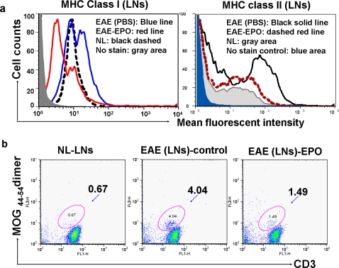
Figure 2. In vivo effect of EPO treatment on MHC expression and number of MOG antigen specific T cells in inguinal lymph nodes.
EPO treatment was started 7 days after mice received MOG35–55 immunization. Bilateral draining inguinal lymph nodes (DILNs) were obtained from mice on day 11 (after 3 day treatment with either EPO at 5000 U/kg/day or sham treatment with PBS) and single cell suspensions were prepared. a) EPO treatment down-regulated mononuclear cell MHC class I (left) and class II (right) expression in lymph nodes from EAE mice. b) Less than 0.7% of single cells from normal mouse inguinal lymph nodes were double-positive for MOG44–54 dimer and anti-CD3 antibody. In contrast, about 4% of single cell suspensions from sham treated EAE mice were positive for FITC-CD3 and PE-MOG40–54/H-2Db dimer double staining (middle), whereas EPO treatment reduced in vivo proliferation of MOG-specific T cells back to 1.5% (right).

