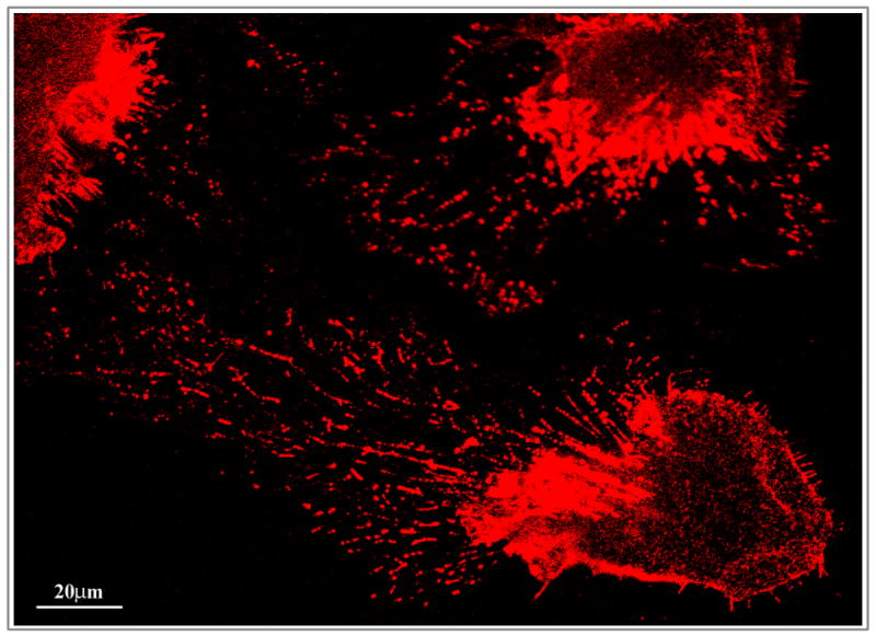
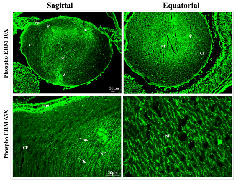
Figure 3.

Distribution of phosphorylated ERM proteins in the neonatal mouse lens and in cultured mouse lens epithelial cells. A. To determine the distribution pattern of activated (phosphorylated) ERM proteins in the lens, P1 mouse lens cryosections (sagittal and equatorial planes) were immunostained with polyclonal antibody raised against phospho-specific ERM protein in conjunction with Alexa Fluor 488 conjugated secondary antibody. Immunofluorescence images were captured using confocal microscopy at 10x (upper panels) and 63X (lower panels) magnification. Arrows and arrowheads indicate staining at the lens fiber cell lateral membrane and lens sutures, respectively. Asterisks in upper panels depict the area that is shown at higher magnification in lower panels. Epi: Epithelium, CF: Cortical fibers and NF: Nuclear fibers. B. Distribution of phospho-ERMs in the cultured mouse lens epithelial cells. The cell apical region and cell surface protrusions including filopodia and microvilli exhibit specific localization of phospho-ERM proteins (red immunofluorescence staining).

