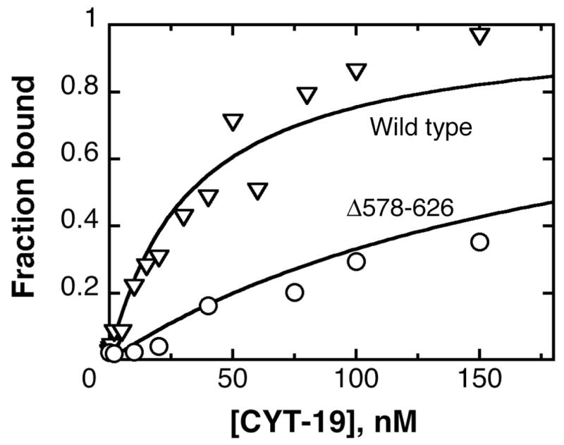
Fig. 5.

Equilibrium binding of Δ578–626 to the Tetrahymena ribozyme, as monitored by nitrocellulose filter binding. The data shown for the Δ578–626 protein (○) gave a Kd value of 200 nM, and the data for the wild-type CYT-19 protein, from an experiment performed side-by-side, (∇) gave a Kd value of 30 nM. The experiments shown did not include added nucleotides; analogous experiments in the presence of 2 mM ADP-Mg2+ or 2 mM AMP-PNP-Mg2+ gave indistinguishable results for both the wild-type and Δ578–626 proteins.
