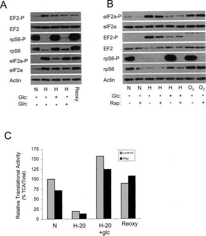
FIGURE 5.
Glucose reverses the effect of hypoxia on the phosphorylation status on rpS6 and eIF2α. (A) Cells were maintained in complete media (*) under normoxic (N) conditions or placed in a hypoxic chamber for 20 h (H). One hour before harvesting, glucose (glc) or glutamine (gln) was added to the media. As a control, hypoxic cells were returned to a normoxic environment for 1 h (Reoxy). Cells were harvested and the phosphorylation status of eIF2α, rpS6, and EF2 was determined by Western analysis. Actin was used as a loading control. (B) Cells were treated with rapamycin and the effect on the phosphorylation of eIF2α, rpS6, and EF2 was analyzed in cells exposed to normoxia (N), hypoxia (H) for 20 h, or cells exposed to hypoxia for 20 h followed by glucose addition for 1 h. Additionally one set of hypoxic cells was returned to normoxic conditions for 1 h (O2). Actin was used as a loading control. (C) The translational activity was measured in PC3 cells grown under normoxic (N) and hypoxic (H-20) conditions in the presence or absence of rapamycin (20 ng/mL). After 20 h of hypoxic treatment, glucose was supplemented into the media (H-20 + glc) for 1 h or returned to normoxic conditions for 1 h (Reoxy) prior to analysis. The translational activity in the normoxic sample without rapamycin was set to 100%. The black bar represents rapamycin treatment (rap) and the gray bar represents untreated cells (control).

