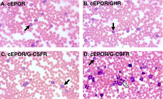
Figure 3.
Constitutive chimera function and expansion of blood cell lineages in the periphery. Peripheral blood smears from various animals were visualized by Wright–Giemsa staining, and representative photomicrographs are presented. (A) Animal transduced with cEPOR. Arrow, reticulocyte. (B) Animal transduced with cEPOR/GHR. Arrow, reticulocyte. (C) Representative animal transduced with cEPOR/G-CSFR and exhibiting marked elevation in hematocrit. Arrow, reticulocyte. (D) Representative animal transduced with cEPOR/G-CSFR and exhibiting marked anemia. Solid arrow, reticulocyte; open arrow, neutrophil.

