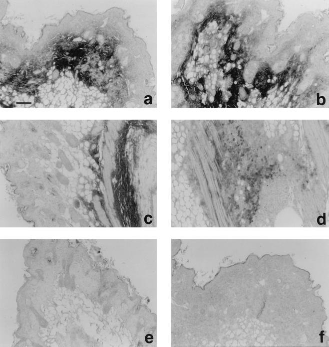
Figure 5.
Immunohistochemical staining of CBEGF and rat EGF injected subcutaneously into nude mice. Immunolocalization was performed on 5-μm paraffin sections by the streptoavidin-biotin-alkaline phosphatase complex technique. Affinity-purified anti-rat EGF antibodies (a-e) or preimmune rabbit serum (f) was used as the primary antibodies. (a, b and f) 5 days after injection of CBEGF (50 μg). (c and d) 10 days after injection of CBEGF. (e) 1 day after injection of rat EGF (10 μg). (Bar, 100 μm.)

