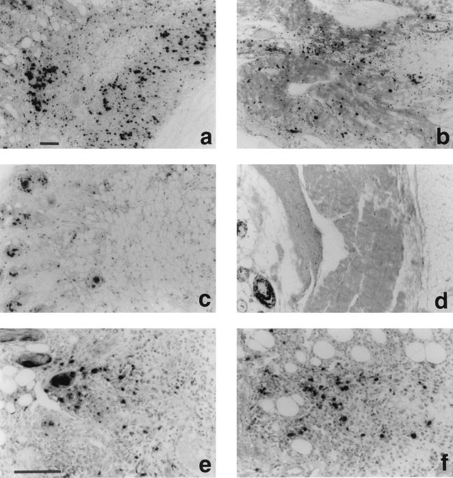
Figure 6.
Detection of S-phase cells as BrdU incorporation and immunohistochemical staining of CBFGF in subcutaneous tissue of nude mice injected with CBFGF and human bFGF. The animals received an i.p. injection of BrdU (10 mg/100 g body weight) 24 h before death. Immunolocalization was performed on 5-μm paraffin sections by the streptoavidin-biotin-alkaline phosphatase complex technique using anti-BrdU mAbs (a-d) or anti-human bFGF antibodies (e and f) as the primary antibodies. (a, e, and f) 5 days after injection of CBFGF (50 μg). (b) 7 days after injection of CBFGF. (c and d) 5 days and 7 days, respectively, after injection of human bFGF (20 μg). (Bars, 100 μm.)

