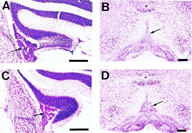
Figure 5.
Extents of floccular and inferior olivary lesions. (A) Coronal section in the middle part of the rostrocaudal extent of the lesioned flocculus, ipsilateral to the observed eye, 7 days after local injection of 0.2 μl of 0.1% ibotenic acid. Note marked cell loss about 60% in granular and Purkinje cell layers. (B) Coronal section of the lesioned inferior olivary nuclei 21 days after i.p. injection of 500 mg/kg of 3-AP followed by 500 mg/kg of nicotinamide. The loss and degeneration of the neurons in the inferior olivary nuclei involving the medial accessory nuclei cell groups are clearly seen. (C) and (D) The intact flocculus and inferior olivary nuclei, respectively. Arrows in A and C indicate choroidal plexus of the IVth ventricle. Arrows in B and D indicate dorsal cap of Kooy. (Bars, 250 μm.)

