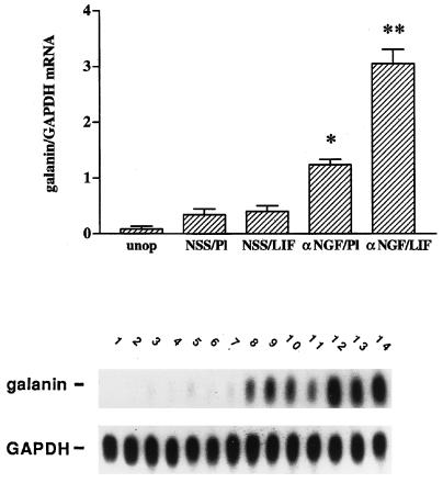
Figure 1.
αNGF treatment alters responsiveness to LIF in intact sympathetic neurons in vivo. Messenger RNA levels were measured by northern blot analysis of SCG (20) that were either untreated (unop), or treated with NSS and a placebo pellet (NSS/Pl), NSS and a LIF-containing slow-release pellet (NSS/LIF), αNGF and a placebo pellet (αNGF/Pl), or αNGF and a LIF pellet (αNGF/LIF). Treatment with antiserum or control serum was for three days, while with LIF pellets or control pellets was for 2 days. In the NSS/Pl group, there was only a small increase in galanin mRNA levels over the unop group. This increase is most likely due to a small amount of neural damage occurring during the process of desheathing the SCG before pellet placement. Levels of galanin mRNA in the NSS/LIF group were not different from those in the NSS/Pl group. There was a 3.5-fold increase in galanin mRNA in the αNGF/Pl group as compared with NSS/Pl treatment. The αNGF/LIF group showed the highest levels of galanin mRNA in this experiment, being 2.3-fold higher than the αNGF/Pl group. Levels of galanin mRNA are expressed as a ratio of the intensity of the galanin band compared with that of the band corresponding to GAPDH. The bar graph represents the means ± SEM of data from five or six samples per group with 2 SCG per sample. The autoradiograph is from one representative experiment and contains multiple lanes of the groups described above. Lanes 1 and 2, unop; lanes 3–5, NSS/Pl; lanes 6–8, NSS/LIF; lanes 9–11, αNGF/Pl; and lanes 12–14, αNGF/LIF. After the blot was probed for galanin, it was stripped and reprobed for GAPDH. *, P < 0.0001 compared with NSS/Pl. **, P < 0.0001 compared with either NSS/LIF or αNGF/Pl.

