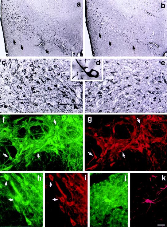
Figure 3.
Predominant expression of D2S protein in the substantia nigra (arrows) as shown with D2S (a, c, d) and D2L (b, e) antibodies. Double labeling immunofluorescence analysis shows colocalization of D2S (FITC, green) and TH immunoreactivity (Cy3, red) in dopaminergic neurons of substantia nigra (f, g), ventral tegmental area (h, i), and retrorubral field (j, k). (Scale bar: a and b, 400 μm; c, 50 μm; d, 11 μm; e, 50 μm; f–k, 50 μm.)

