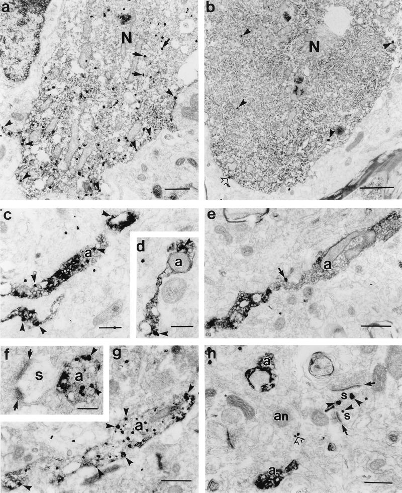
Figure 4.
Electron micrographs of double labeled sections showing differential D2S and D2L receptor expression in dopaminergic neurons and axons. Dopaminergic markers, TH or dopamine transporter (DAT), are labeled by immunoperoxidase reaction and receptors by immunogold method. In dopaminergic neurons of substantia nigra, note the significantly higher number of immunogold particles (arrowheads) in the D2S (a) labeled cell body than D2L (b). D2S immunogold particles are frequently distributed at the cytoplasmic face of plasma membranes (arrowheads) and rough endoplasmic reticulum (arrows). D2L immunogold particles are only occasionally associated with plasma membranes (open arrow in b). TH immunolabeled axons (a) exhibit numerous D2S (c, d) immunogold particles (arrowheads), whereas D2L (e, h) immunoreactivity is not detected. In e, the immunogold particle is outside the TH-labeled axon (arrow). In h, D2L immunogold particles are localized along the cytoplasmic side of plasma membranes in dendritic spines (s; arrowheads), which receive asymmetric synapses (arrows). TH immunonegative axon (an) expresses D2L immunoreactivity (open arrow). (f–g) Dopaminergic axons in the area 6 labeled with DAT are strongly D2S positive. In f, D2S-labeled axon is apposed to the spine receiving an asymmetric input (arrows). N, nucleus. (Scale bars: a, 0.5 μm; b, 1 μm; c and d, 0.2 μm; e, 0.5 μm; f, 0.2 μm; g, 0.5 μm; h, 0.3 μm.)

