Figure 5.
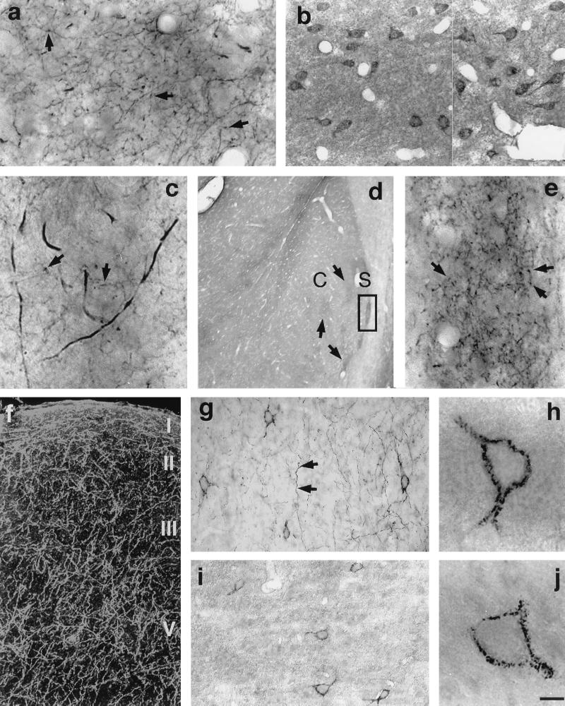
Dense D2S immunopositive fiber network in the projection areas of dopaminergic neurons: striatum (a, c), nucleus accumbens (d, e), and cerebral cortex area 6 (f, darkfield micrograph; g). Immunopositive fibers bear numerous varicosities (arrows). (e) High-magnification micrograph depicted from square shown in d. D2L-positive neurons in striatum (b) and cerebral cortex area 6 (i, j) are shown. (g, h) Neurons labeled with D2S antibody in the area 6 of cerebral cortex. C, core; S, shell. (Scale bar: a and b, 25 μm; c, 8 μm; d, 400 μm; e, 25 μm; f, 80 μm; g and i, 67 μm; h and j, 10 μm.)

