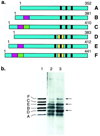
Figure 4.
(a) Schematic representation of the six human brain tau isoforms, with the alternatively spliced exons shown in red (exon 2), green (exon 3), and yellow (exon 10). The microtubule-binding repeats are indicated by black bars. (b) Immunoblots of dephosphorylated soluble tau protein from the frontal cortex of a control subject (lane 2) and a patient with familial MSTD (lane 3) using anti-tau serum BR133. Similar results were obtained with anti-tau serum BR134. Six tau isoforms are present in lanes 2 and 3. They align with the six recombinant human brain tau isoforms (lane 1). In the frontal cortex from the familial MSTD patient, tau isoforms with four repeats (isoforms D, E, and F) are more abundant and tau isoforms with three repeats (isoforms A, B, and C) are less abundant than in frontal cortex from the control. Arrows indicate the positions of tau isoforms with four repeats.

