
Figure 8.
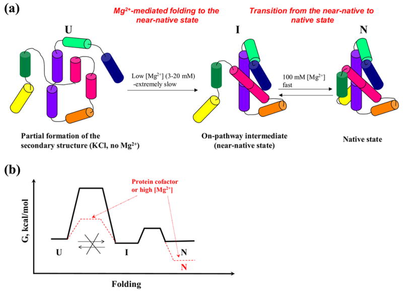
(a) A scheme illustrating folding pathway of the ai5γ-derived ribozyme at 30 °C. (b) A scheme of a hypothetical free energy diagram for the ribozyme folding to the near-native state and native states at low magnesium (solid black lines). Hypothetical changes in free energy landscape upon adding high concentrations of magnesium or protein co-factors are shown in red.