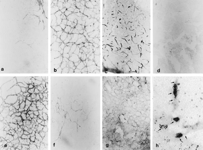
Figure 2.
Depiction of fetal cortical explants after VEGF treatments. Immunostaining for laminin (a–e) shows only a few vascular profiles in control explant (a) whereas the 25-ng dose produces a significant angiogenic network in fetal (b) and newborn (e) (nickel intensification) explants. At the 100-ng dose angiogenesis is tapered off (c) (nickel intensification) and addition of neutralizing antibody to VEGF abolishes all angiogenic effects (d). Immunoexpression of GLUT-1 depicts substantially fewer vessel profiles than laminin and is most prominent at the 1-ng dose (f) whereas patchy immunoexpression of the Flt-1 receptor is found on most vessels at the 25-ng dose (g). After [3H]thymidine administration, anti-laminin (+) vascular profiles are labeled in a 7-μm paraffin section (arrows in h). All micrographs except h are identical magnification.

