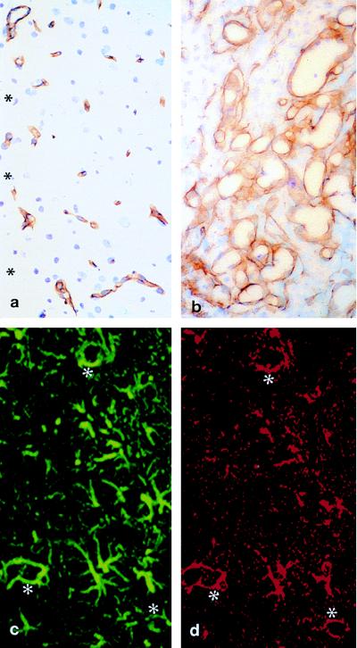
Figure 7.
Minipump infusions of VEGF into the adult cortex. Control (saline infusion) (a) shows no measurable changes in the cerebrovasculature as shown by laminin immunostaining along the wound edge (∗). After VEGF infusion a robust angiogenic effect produces a substantial number of tightly packed, dilated vessels (b). a and b are at identical magnification. The VEGF flt-1 receptor is expressed by reactive astrocytes after VEGF infusion. Laser confocal microscopy shows that immunoexpression of GFAP (c) near the infusion site mostly colocalizes with the flt-1 receptor (d). Note the dual labeling of perivascular astroglia (∗ in c and d).

