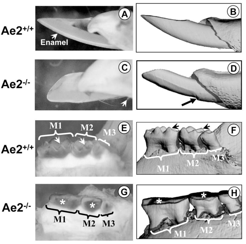
Figure 1A – H.

Fig. 1A is a macro-photograph of an erupted Ae2a,b+/+ incisor and Fig. 1B the μCT image of the same tooth. In Fig. 1A, the enamel can be clearly seen. Fig. 1C is a macro-photograph of an Ae2a,b−/− incisor. Note that there is virtually no enamel present, only some remnants (white arrows). The corresponding μCT image of the same tooth is shown in Fig. 1D. The black arrow shows the eroded enamel just before it enters the oral cavity. The morphology of the Ae2a,b+/+ molars is shown in Figs 1E (macro-photograph) and 1F (μCT), respectively. The cusps of all molars (M1= first, M2= second and M3= third maxillary molars) are sharp (arrows) and are not eroded. The molars from the Ae2a,b−/− specimen (Fig. 1G, macro-photograph and Fig. 1H, the corresponding μCT image of the same tooth) are severely eroded (asterisks).
