
Figure 2. Localization of tubulin PTMs on the α/β-tubulin heterodimer.
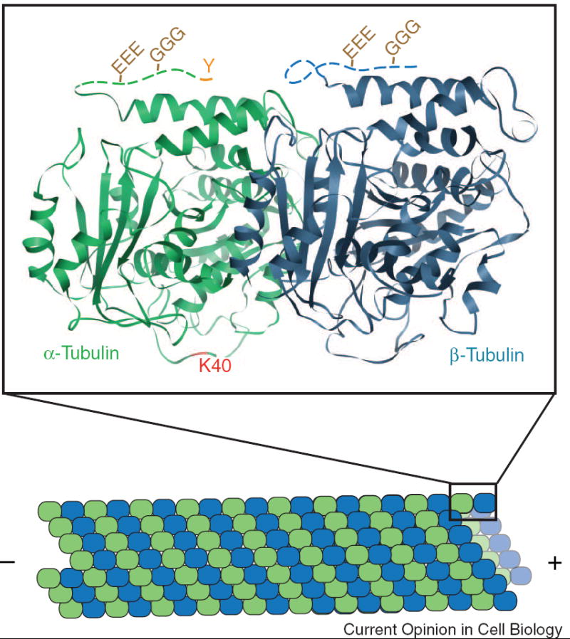
Microtubules are formed by the end-on-end and lateral associations of α- (green) and β- (blue) tubulin heterdimers. The crystal structure (PDB file 1TUB) of one tubulin heterodimer is shown. The CTTs of both α- and β-tubulin, disordered in the structure, are represented in this schematic as dashed lines. Detyrosination entails the removal of the C-terminal tyrosine (Y) from α-tubulin. The resulting detyrosinated α-tubulin is often referred to as Glu tubulin due to the exposed glutamate residue. Polyglutamylation (EEE) and polyglycylation (GGG) occur on the CTTs of α- and β-tubulin. Acetylation of α-tubulin occurs on lysine40 (K40) which is predicated to lie on the luminal face of the microtubule.