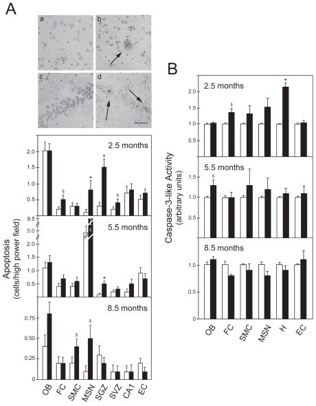
Figure 1.
DNA fragmentation and caspase-3 activation occur early in the frontal cortex and hippocampus of rTg4510 mice. Brain slices were analyzed from both control (white bars) and transgenic animals (black bars) at 2.5, 5.5, and 8.5 months. DNA fragmentation was assessed by the TUNEL assay, and the number of positive cells was counted in the olfactory bulb (OB), frontal cortex (FC), sensorimotor cortex (SMC), medial septal nucleus (MSN), subgranular zone (SGZ), subventricular zone (SVZ), CA1, and entorhinal cortex (EC). Cytosolic proteins were extracted for caspase-3-like activity assays from OB, FC, SMC, MSN, hippocampus (H), and EC of both control and transgenic animals at 2.5, 5.5, and 8.5 months. A: TUNEL staining in control FC (a); transgenic FC (b); control H (c); and transgenic H (d) at 2.5 months (top) and number of apoptotic cells (bottom). Apoptotic nuclei were identified by a condensed nucleus with dark staining (arrows). B: DEVD-specific caspase activity. The results are expressed as mean ± SEM of at least three different experiments. *P < 0.01 and §P < 0.05 from control animals. Scale bar: 100 μm.

