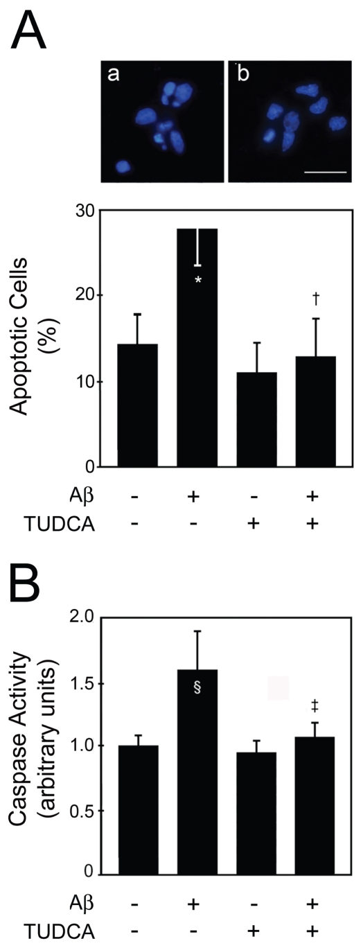
Figure 4.
TUDCA inhibits apoptosis and caspase-3 activation in primary cortical neurons incubated with fibrillar Aβ1–42. Cells were incubated with 20 μM fibrillar Aβ1–42, or no addition (control), ± 100 μM TUDCA for 24 h. In coincubation experiments, TUDCA was added 12 h prior to incubation with Aβ1–42. Cells were fixed and stained for microscopy assessment of apoptosis. Cytosolic proteins were extracted for caspase-3-like activity assays. A: Fluorescence microscopy of Hoechst staining in controls (a); and in cells exposed to Aβ1–42 (b) (top) and percentage of apoptosis (bottom). Apoptotic nuclei were identified by condensed chromatin as well as nuclear fragmentation. B): DEVD-specific caspase activity. The results are expressed as mean ± SEM of at least three different experiments. *P < 0.01 and §P < 0.05 from control; ‡P < 0.01 and †P < 0.05 from Aβ. Scale bar: 15 μm.

