
Figure 1.
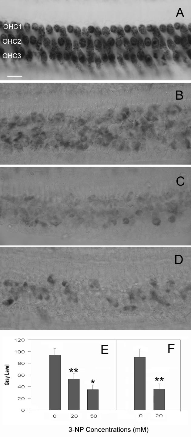
Comparison of the levels of SDH reduction. A: SDH staining in a normal control cochlea that received only the artificial perilymph solution. The dark staining in OHCs indicates strong SDH activity. B: SDH activity following the treatment of 20 mM of 3-NP on the round window. Notice a significant reduction of SDH staining. C: SDH staining after the treatment of 50 mM of 3-NP on the round window. SDH activity appears further reduced. D: SDH activity following cochlear perfusion of the 3-NP solution (20 mM). SDH staining is substantially reduced to a level similar to that induced by the round window application of 3-NP at 50 mM. E: Comparison of the average gray levels of SDH staining among the cochleae treated with the artificial perilymph solution (control), 20 mM or 50 mM of 3-NP solutions on the round window. F: The comparison of the gray levels of SDH staining between the 3-NP perfused ears and the artificial perilymph-perfused control ears. OHC1, OHC2, and OHC3 represent the first, second and third row of OHCs respectively. * indicates p < .05 and ** indicates p < .01. Bar: 20μm.