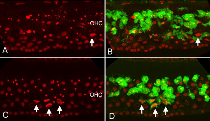
Figure 4.
Double staining of PI and caspase-3 activity in a 3-NP treated ear and a control ear without the 3-NP treatment following exposure to the noise. A: Nuclear morphology of OHCs in the perilymph-treated control ear. B: The same cochlear section showing both the nuclear morphology and the caspase-3 fluorescence (green). Notice that strong caspase-3 activity is observed in the OHCs exhibiting nuclear condensation or fragmentation. However, there is no caspase-3 activity in the dying cells with swollen nuclei (indicated by an arrow in A and B). C and D: the PI fluorescence (C) and PI plus caspase-3 (D) in a 3-NP-treated cochlea. Similar to the perilymph-treated control cochlea (see A and B), the 3-NP treated cochlea shows caspase-3 activity in all the OHCs having condensed nuclei. However, certain OHCs with necrotic nuclei also exhibit caspase-3 activity (arrows). The labels of OHC in A and B indicate the OHC region.

