
Figure 3.
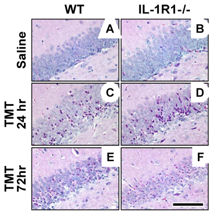
Representative hematoxylin and eosin (H&E) staining in the suprapyramidal blade of the dentate gyrus in wildtype (WT) and IL-1R1−/− mice under control conditions (A, B) and following TMT treatment (2.0 mg/kg, i.p., C–F). No differences were observed between the groups at basal level (A, B), or 24 hrs following TMT (C, D). Severity scoring of the insult based on number and location of eosin positive cells verified comparable levels of damage in WT (C, 2.25 ± 0.46) and IL-1R1−/− (D, 2.0 ± 0.58) mice at 24 hrs. Examining WT and IL-1R1−/− mice at 72 hrs post-TMT (E, F) resulted in each group reaching a severity score of 4, indicative of granule cell loss. Scale bar = 100µm.