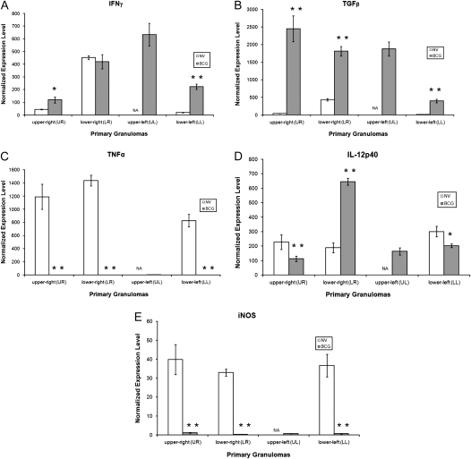
Figure 2.
Cytokine mRNA expression of primary granulomas from guinea pigs aerosol- challenged with Mycobacterium tuberculosis. Expression of (A) IFN-γ mRNA, (B) TGF-β mRNA, (C) TNF-α mRNA, (D) IL-12p40 mRNA, and (E) iNOS mRNA were measured from granulomas microdissected from the four lung lobes (upper right, lower right, upper left, lower left) of nonvaccinated (open bars) and BCG-vaccinated (shaded bars) guinea pigs 6 weeks after virulent challenge. Values for threshold cycle (Ct) were converted to “expression levels” to allow for fold comparisons between samples, expression level = 2(40-Ct). Data were normalized to HPRT rRNA followed by values derived from nongranulomatous tissues and expressed as means ± SEM (n = 8–10 capfuls of microdissected tissue from various regions of lung lobes). Large, necrotic, primary lesions were not found in the upper left lung lobe of nonvaccinated animals. Asterisks indicate significant (*P < 0.05) or highly significant (**P < 0.01) differences found between granulomas from nonvaccinated and BCG-vaccinated guinea pigs.

