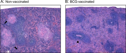
Figure 6.
Spleens from (A) nonvaccinated and (B) BCG-vaccinated guinea pigs at 6 weeks after infection with low-dose M. tuberculosis were fixed in buffered formalin and stained with H&E. The arrows in A indicate the presence of splenic granulomas surrounding the lymphoid follicles of nonvaccinated guinea pigs. The arrowhead in B indicates the white pulp zone, located around a central arteriole, in the spleens of BCG-vaccinated guinea pigs. Total magnifications for both panels: ×500.

