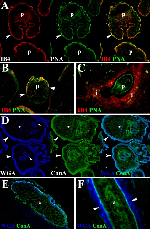
Figure 4. Release of tegument material in mice intracranially infected with M. corti.
(A) Peritoneal M. corti (p) stained in the tegument (arrowheads) with IB4 and PNA. 200× (B) Parasite (p) invading nervous tissue 2 DPI. Tegument in contact with host tissue (arrowheads) has lost IB4 and PNA GC ligands. 200× (C) After 3 weeks of IC infection the parasite (p) is IB4 negative but the infiltrating cells are IB4 positive. PNA bound GCs are found in the whole parasite. 200× (D) M. corti tegument (large arrowheads) stained with WGA and ConA. Metacestode parenchyma (asterisks) is mostly stained with ConA. Scolex tegument (small arrowheads) is also stained with both lectins. 200× (E) M. corti metacestode in leptomeninges after 3 DPI. ConA binding material is present in tegument and parenchyma (asterisk), WGA binding material is released from the tegument (arrowheads). 200× (F) At 3 weeks of IC infection M. corti remains positive for ConA and WGA in the tegument, but a portion of the WGA binding material (arrowheads) continues to disassociate from the parasite 400×.

