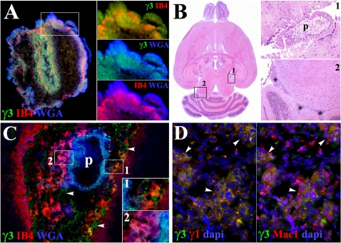
Figure 5. Host cells phagocytose M. corti GCs during the course of murine NCC.
(A) γ3, IB4 and WGA staining colocalize in tegument and parenchyma of IP maintained parasite after multiple washes with HBSS (400×). Distinct combinations of lectin and γ3 staining in the tegument are shown in the right panels (B) H&E staining of mouse brain 3 wks PI (10×). Inset 1 (200×) is showing parasite (p) located in internal leptomeninges similar to that in C. Inset 2 (50×) shows perivascular infiltrates (asterisks) located distal to parasites, details of such infiltrates are shown in D (50×) (C) M. corti (asterisk) stained with WGA and γ3, but not IB4 in internal leptomeninges 3 wks PI. Infiltrating cells are double positive for IB4 and MCSγ3 (inset 1) and for WGA and IB4 (inset 2). Free parasite material is also detected (arrowheads). 200×. Inset 1 and 2 are 2.5 times magnified. (D) Inflammatory infiltrates (arrowheads) in internal leptomeninges showing γ3 and γ1 staining. Panel on the right shows that parasite material labeled by γ3 and γ1 is present in Mac1+ cells 3 wks PI. 400×.

