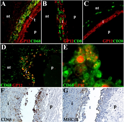
Figure 7. Host cells taking up T. solium GP12.
Different leukocyte populations were detected with monoclonal antibodies and streptavidin-Alexa 488 (green), streptavidin-HRP (brown). Anti-GP12 polyclonal antibody was labeled with Alexa 546 (red). Samples A–C are from non-granulomatous lesions and D–G from samples with granuloma formation. Nervous tissue (nt). (A) Anti-GP12 strongly labels the whole parasite (p) tegument (t) and the macrophages (CD68) present in the infiltrate (i) 400×. (B) CD8 T cells do not colocalize with anti-GP12 in the i. The p represents the space where the parasite was lodged. 400× (C) Anti-GP12 stains some cells in the i, but no B cells (CD20). 400× (D) p surrounded by i showing macrophages co-localizing with anti-GP12 (orange-yellow color). 400× (E) Epithelioid histiocytes and giant cells showing anti-GP12 staining in most of the vacuoles present in their cytoplasm. 1000× (F) Macrophages and multinucleated giant cells (dotted line) surrounded the p and located in i forming granuloma. 400× (G) Low to undetectable expression of MHC-II in macrophages (dotted line) adjacent to the p. Moderate MHC-II expression is detected in the i. 400×.

