
Figure 2.
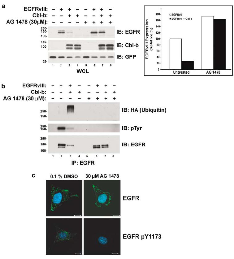
Inhibition of the TK activity of the EGFRvIII prevents its downregulation and ubiquitination by Cbl-b. CHO cells were transfected with the EGFRvIII, HA-epitope-tagged ubiquitin, and Cbl-b as indicated. All transfections were balanced with empty vector controls; GFP was used as a transfection control. Following transfection, cells were grown to 70% confluence and incubated overnight with the vehicle (0.1% DMSO) or 30 μm AG 1478. (a) Whole-cell lysates (WCL) or (b) EGFR immunoprecipitates (IP) were immunoblotted (IB) for the EGFR, Cbl-b, GFP, ubiquitin (with anti-HA), or phosphotyrosine (pTyr) as indicated to the right of the blots. The bar graph shows the EGFRvIII protein levels in the WCL blots in panel a quantified by densitometry. All values, expressed as the percentage of the amount of EGFRvIII protein in the control transfection, are adjusted for GFP levels. Molecular weight standards (kDa) appear to the left of the panels. (c) EGFRvIII-expressing NR-6m cells were incubated with 100 μg/ml cycloheximide (to inhibit new protein synthesis) and either 0.1% DMSO or 30 μm AG 1478, fixed, and stained for the EGFR or activated (pY1173) EGFR as indicated (green). Cells were counterstained with DAPI (blue). Each panel is a representative mid-level confocal slice. Bar=10 μm.