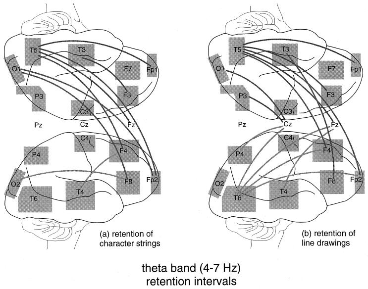
Figure 3.
Enhanced coherence in the θ range (4–7 Hz) during preception and retention intervals. Connections between electrode sites represent significant increases of coherence above control (P < 0.05 or better). The significance level was evaluated by applying paired Wilcoxon tests to group results. The shaded areas indicate the range of positions of individual electrodes as determined in an MRI study (24). Note that the occipital electrodes (O1, O2) are placed not over primary visual areas, but closer to the parieto-temporo-occipital association region. (a) During retention of character strings in memory (task I), enhanced coherence appeared between prefrontal and posterior cortex. In posterior cortex, the left hemisphere was predominantly involved. (b) Coherence-increases during retention of abstract line drawings in memory (task II). Patterns of enhanced coherence were similar to those in the verbal memory task a, although more connections appeared in the right hemisphere.

