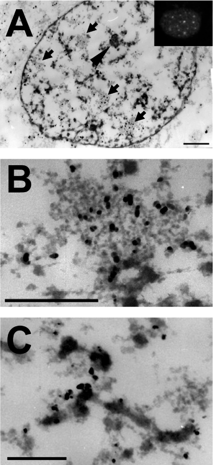
Figure 2. Ultrastructural immunolocalization of Hsp25 in the nuclei of heat-shocked Rat-1 fibroblasts.
To identify the intranuclear localization of rodent Hsp25 after heating at the ultrastructural level, heated (30 min at 43 °C) Rat-1 cells were briefly lysed in TEA buffer containing Mg2+ prior to standard fixation in paraformaldehyde. After immunolabelling with a primary anti-Hsp25 antibody and a secondary goat anti-rabbit 5 nm gold-labelled antibody, ultrathin sections of the cells were processed for transmission electron microscopy. (A) Entire cell nucleus of a cell heat shocked for 30 min. Arrows indicate IGCs and the arrowhead indicates the nucleolus. The inset represents a typical nucleus of a similarly treated cell, stained for immunofluorescence. Bar=1 μm. (B) Enlarged image of an IGC clearly enriched with the Hsp27 label. Bar=1 μm. (C) Enlarged fragment of a perichromatin fibre, containing the label along its periphery. Bar=1 μm.

