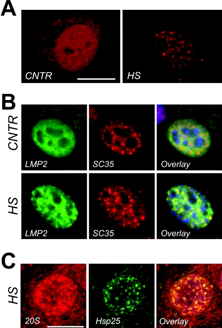
Figure 6. Association between nuclear granules of Hsp25, nuclear speckles and the 20S proteasomal machine.
(A) Confocal images of cell nuclei decorated with anti-20S proteasomal antibodies in the nuclei of cells before (CNTR) and 60 min after a 30 min heat shock at 43 °C (HS). Bar=10 μm. (B) Multicolour channel images of nuclei, expressing GFP-labelled 20S core subunit LMP2 before (panel labelled CNTR) and after a 30-min heat shock at 43 °C (panel labelled HS). Each horizontal series represent the same nucleus in which epigenetical LMP2–GFP (LMP: green) and endogenous SC35 (SC35: red) are revealed and overlaid (Overlay: co-localization is evident by the presence of yellow colour). 20S proteasomes (LMP) become concentrated in nuclear speckles (SC35) after heat shock. Bar=5 μm. (C) Confocal images of heat-shock cells immunostained with the anti-Hsp25 antibody (Hsp25: green) and the anti-20S proteasome antibody (20S: red). Co-localization is evident in the overlay image (Overlay: yellow). On the overlaid images nuclear outline is indicated with blue (DAPI staining). Bar=10 μm.

