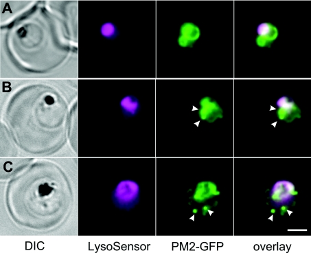
Figure 7. Labelling of 3D7-PM2–GFP transfectants with LysoSensor Blue.
Mixed-stage transfected parasites were incubated with the pH probe, LysoSensor Blue. Shown are bright field, LysoSensor Blue, GFP and merged GFP/LysoSensor Blue images. The LysoSensor Blue fluorescence is largely associated with the DV, while the GFP chimaera is also present in cytostomal vesicles (arrows) that do not stain with the LysoSensor Blue. Scale bar, 2 μm.

