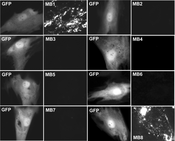
Figure 4.
Fluorescence signal from nine different MBs targeting BMP-4 mRNA and from GFP HDF cells 2 days after adenovirus infection. Left and right panels display respectively the epifluorescence images of GFP expression and signal from MBs targeting BMP-4 mRNA in the same cell. The same exposure times were used for GFP and MB imaging. The results showed clearly that only MB1 and MB8 gave high signal level, indicating good target accessibility, whereas all other MBs (MB2–MB7) have poor target accessibility, as indicated by the very weak signal levels.

