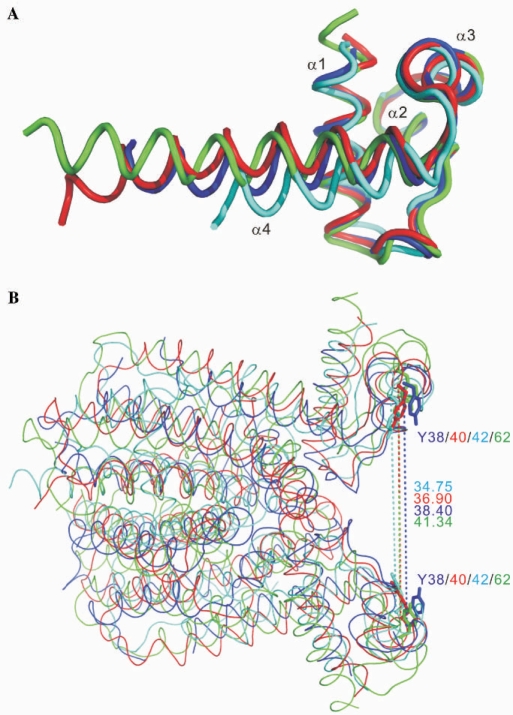
Figure 4.
Structural homologues with distinct DNA recognition modes. (A) Superimposition of the three-helix bundle DNA-binding domains. The similarity between IcaR (blue), TetR (cyan), QacR (red) and EthR (green) shows a high degree of structural homology within the DNA-binding domains of these proteins. (B) Comparison of the dimer structures. The dimers of IcaR, TetR, QacR and EthR are superimposed and colored as in (A). The distances between the two Cα atoms of Tyr38 in the IcaR dimer and the equivalents in TetR, QacR and EthR, which are supposed to interact with the DNA bases in two adjacent major grooves, have a narrow range of 35–41 Å.

