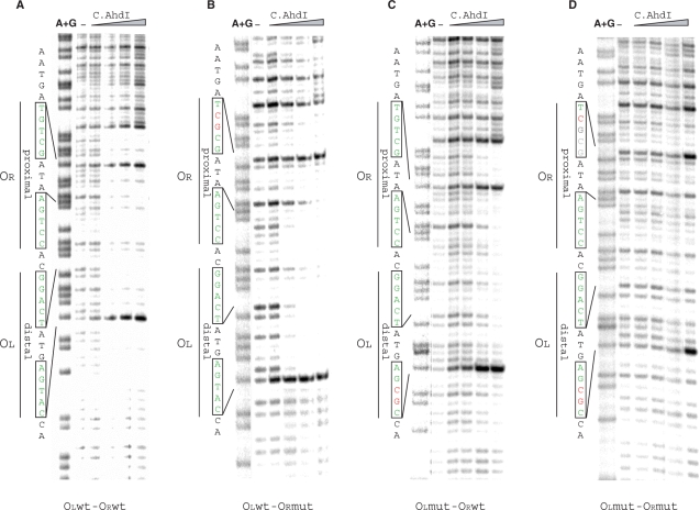
Figure 4.
DNase I footprinting of C.AhdI complexes on wild-type and mutant ahdICR promoters. 32P-end labelled (bottom strand) ahdICR promoter-containing fragments (22.5 nM) were combined with increasing concentrations (0–188 nM) of C.AhdI and treated with DNase I. Two sets of inverted repeats are indicated at the left of each panel.

