Figure 3.
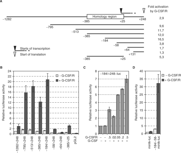
The MAD1 promoter is activated by G-CSF. (A) Schematic representation of the MAD1 promoter fragments that were cloned into pGL2. The homology region that shows high sequence conservation between human, mouse and rat is indicated. Furthermore, the major start sites of transcription and the position of the ATG are shown. The fold activation in response to G-CSF/G-CSFR of each promoter construct is indicated. These numbers are derived from the data shown in (B). (B) RK13 cells were transiently transfected with plasmids expressing the G-CSFR (0.5 µg) and the indicated promoter-luciferase reporter gene construct (1 µg) and treated with G-CSF (G-CSF/R). Luciferase activities were standardized with co-expressed β-galactosidase. The mean values and standard deviations of three independent experiments performed in duplicates are displayed. (C) The experiments were performed as in panel B with the indicated reporter construct and increasing concentrations of the G-CSFR expressing plasmid. (D) The experimental design was as in panel B. The –184 to –58 fragment of the MAD1 promoter was cloned 5′ of the minimal thymidin kinase promoter (mintk)-luciferase reporter gene construct.

