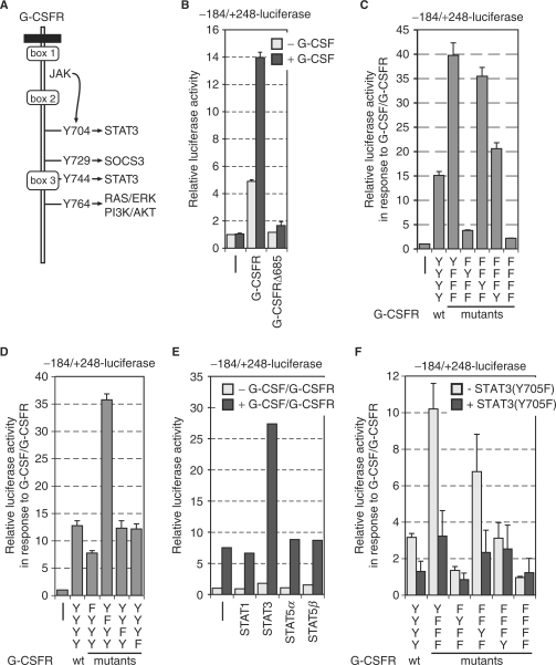
Figure 5.
STAT3 is important to activate the MAD1 promoter. (A) Schematic representation of the cytoplasmic portion of the G-CSF receptor (G-CSFR). The conserved boxes 1–3 and the four tyrosine residues that are known to be phosphorylated in response to G-CSF are indicated. The signal transduction pathways that are connected to the individual tyrosine residues are given. (B) RK13 cells were cotransfected with the –184 to +248 MAD1 promoter luciferase reporter construct and expression plasmids for the indicated wild-type G-CSFR or deletion mutants. The cells were stimulated with G-CSF prior to harvesting and measuring luciferase and β-galactosidase activity. A typical experiment performed in triplicates is shown. (C and D) The experimental design was as in (B). G-CSFR mutants with the indicated changes of either individual tyrosine residues or combination of tyrosines were analyzed. (E) The transfections were performed as in panel B with the additional co-expression of different STAT factors as indicated. A typical experiment performed in duplicates is shown. (F) The experiment was done as in panel B. A dominant negative version of STAT3 [STAT3(Y705F)] was co-expressed as indicated.

