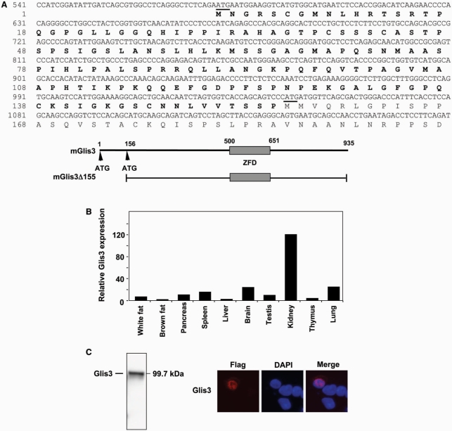
Figure 1.
(A) Nucleotide and amino acid sequence of Glis3. The new extended N-terminal sequence of Glis3 is indicated in bold; only the 5′-end of Glis3 is shown. The start codons are underlined; the first start codon belongs to full-length Glis3, the second one to the previously reported Glis3(ΔN155) (9). A schematic view of Glis3 is shown below the sequence. ZFD indicates zinc finger domain. (B) Expression of full-length Glis3 mRNA in several mouse tissues. Total RNA isolated from various adult tissues was examined by real time QRT-PCR analysis using primers specific for the 5′-UTR of full-length Glis3. (C) The full-length Glis3 localizes to the nucleus. p3×FLAG-CMV-Glis3 plasmid was transfected in C3H10T1/2 cells and after 48 h incubation proteins were isolated and examined by western blot analysis using anti-FLAG M2 antibody. Subcellular localization of FLAG-Glis3 was examined by confocal microscopy with anti-FLAG M2 and Alexa Fluor 594 antibodies; nuclei were identified by DAPI staining.

