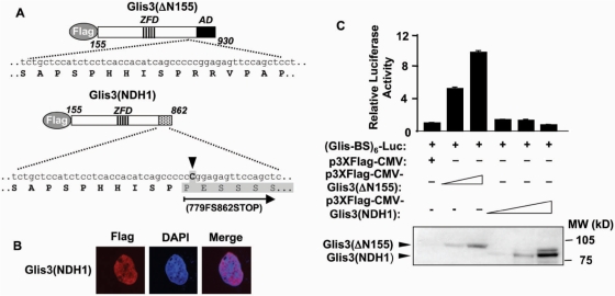
Figure 9.
Glis3(NDH1) localized to the nucleus, but lost its transactivation activity. (A) Schematic view of Glis3(Δ155) and Glis3(NDH1). ZDF and AD are indicated. An insertion of C (arrow head) in the Glis3 gene (NDH1) is linked to the most severe type of NDH. This insertion results in a frame shift at 779 and a stop codon at 862 (779FS862STOP) as indicated (shaded box). (B) Glis3(NDH1) localizes to the nucleus. HEK293T cells were transfected with p3×FLAG-CMV-Glis3(NDH1) and subcellular localization was examined by confocal microscopy with anti-FLAG M2 and Alexa Fluor 594 antibodies; nuclei were identified by DAPI staining. (C) Glis3(NDH1) lacks transcriptional activity. HEK293T cells were co-transfected with p3×FLAG-CMV-Glis3(ΔN155), p3×FLAG-CMV-Glis3(NDH1), pCMVβ and (Glis-BS)6-LUC as indicated. After 30 h, cells were assayed for luciferase (LUC) and β-galactosidase activity. The relative LUC reporter activity was calculated and plotted. The expression of Glis3(ΔN155) and Glis3(NDH1) proteins was examined by western blot analysis using anti-FLAG antibody (lower panel).

