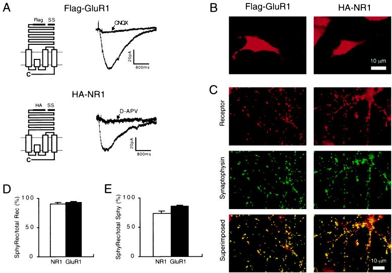
Figure 1.
Epitope-tagged GluR1 and NR1 form functional channels and are targeted to synaptic membranes. (A) (Left) Diagram of the epitope-tagged GluR1 (Upper) and NR1 (Lower). SS, signal sequence. (Right) The inward currents generated by application of glutamate to HEK293 cells expressing Flag-GluR1 (Upper) or HA-NR1 with NR2B (Lower). The currents are blocked by the appropriate subtype-specific antagonist. (B) Examples of the receptor distribution that is observed when hippocampal neurons expressing Flag-GluR1 or HA-NR1 are permeabilized before applying the primary receptor antibodies. (C) (Top) Typical receptor distributions when neurons are not permeabilized and stained for Flag-GluR1 or HA-NR1. (Middle) The distribution of synaptophysin puncta in the same fields. (Bottom) The superimposed images illustrate that the vast majority of Flag-GluR1 and HA-NR1 clusters colocalize with synaptophysin. (D) Quantitation of the percentage of Flag-GluR1 and HA-NR1 clusters that colocalize with synaptophysin (n = 20 for each subtype) indicates that the vast majority of Flag-GluR1 and HA-NR1 clusters are at synapses. (E) Quantitation of the percentage of synaptophysin puncta that colocalize with Flag-GluR1 and HA-NR1 (n = 20 for each subtype) indicate that the majority of synapses are excitatory and contain epitope-tagged receptors. The remaining synapses presumably are inhibitory because 10–30% of synaptophysin puncta colocalized with GAD65 (see Fig. 2D). Error bars represent SEM.

