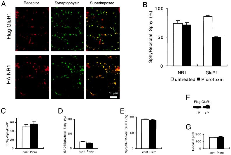
Figure 2.
Increasing activity causes a decrease in synaptic membrane clusters of Flag-GluR1 but not HA-NR1. (A) Example of the staining patterns that are observed after treatment with picrotoxin. Note that many of the synaptophysin puncta do not colocalize with Flag-GluR1. (B) Quantitation of the percentage of synaptophysin puncta that colocalize with Flag-GluR1 or HA-NR1 in untreated and picrotoxin-treated cultures. Picrotoxin had no effect on HA-NR1 but caused a significant reduction in the synaptic localization of surface clusters of Flag-GluR1 (n = 20 for each condition). (C) The average number of synapses per microscopic field, as defined by synaptophysin staining, was not affected by picrotoxin treatment. (D) The percentage of inhibitory synapses, as measured by GAD65 staining, was not affected by picrotoxin treatment. (E) The percentage of Flag-GluR1 clusters that colocalize with synaptophysin was not affected by picrotoxin treatment. (F) Western blot showing that picrotoxin treatment did not change the level of expression of Flag-GluR1. (G) Total Flag-GluR1 immunoreactivity in the somas of permeabilized cells was not affected by picrotoxin treatment, as measured by integration of total receptor immunoreactivity using NIH Image software. Error bars represent SEM.

