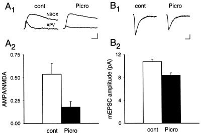
Figure 4.
AMPAR-mediated synaptic currents are decreased by picrotoxin treatment. (A) The ratio of AMPAR- to NMDAR-mediated synaptic currents is decreased in picrotoxin-treated cultures. Examples of the EPSCs (A1) recorded from untreated and picrotoxin-treated cultures and (A2) a summary (n = 5 for untreated cells; n = 6 for picrotoxin-treated group) of the AMPAR-to-NMDAR EPSC ratios obtained from each group. Note that the NMDAR EPSCs were scaled for ease of comparison. [Scale bar represents 25 msec and 15 pA (untreated) or 50 pA (picrotoxin).] (B) The amplitude of mEPSCs is decreased in picrotoxin-treated cultures. Examples of mEPSCs (average of 200–400) (B1) and a summary of all recordings (B2) (n = 6 for control cultures; n = 5 for picrotoxin-treated cultures). (Scale bar represents 10 msec and 2 pA.) Error bars represent SEM.

