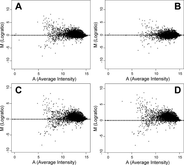
Figure 2.
MA-plots of aCGH data after applying different normalization methods. The log transformed ratios of slide 11 [17] are plotted against the log transformed sum of the green (negative M values; MG1363 signals) and red (positive M values; IL1403 signals) channels. A: non-normalized data. B: grid-based Lowess normalization. C: S-Lowess normalization based on the LCG set obtained from the comparison of L. lactis IL1403 amplicon sequences to the ORFs of three S. pneumoniae strains. D: S-Lowess normalization with a stringent LCG set (99% identity over 100 bp).

