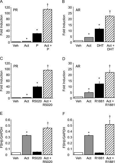
Figure 1.
Progestins and androgens synergize with activin to induce FSHβ gene expression in immortalized gonadotrope and mouse primary pituitary cells. A–D, The −1000FSHβluc reporter plasmid was transiently transfected into LβT2 cells along with 200 ng rat PR (A and C) or AR (B and D). After overnight starvation in serum-free media, the cells were treated for 24 h with vehicle (Veh), 10 ng/μl activin (Act), 100 nm progesterone (P) (A), DHT (B), R5020 (C), R1881 (D), or both activin and the relevant steroid hormone, as indicated. Luciferase activity was normalized to β-galactosidase activity and set relative to the empty reporter vector. Results represent the mean ± sem of at least three independent experiments performed in triplicate and are presented as fold induction of hormone treatment relative to the vehicle control. E and F, Primary pituitary cells were dispersed from 8-wk-old male mice and plated in culture. The cells were treated for 24 h with vehicle (Veh), 10 ng/μl activin (Act), 100 nm R5020 (E), R1881 (F), or both activin and the relevant steroid hormone, as indicated. Total RNA was purified and reverse transcribed, and the level of FSHβ expression was assayed by real-time PCR. In each sample, the amount of FSHβ, calculated from the standard curve, was compared with the amount of GAPDH. The results represent the mean ± sem of FSHβ levels relative to GAPDH performed in triplicate. *, Significant differences from the vehicle-treated control; †, synergistic interaction as defined by a two-way ANOVA (P < 0.05).

