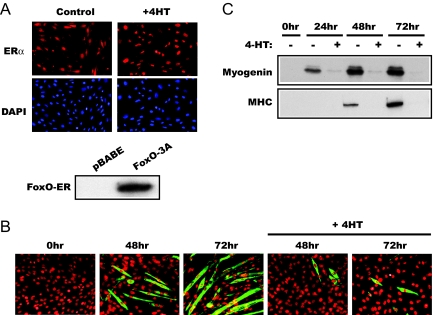
Figure 1.
Active FoxO1 inhibits C2C12 differentiation. A, C2C12 cells stably expressing FoxO1–3A-ER were immunostained using an anti-ER antibody, accompanied by 4′,6-diamidino-2-phenylindole nuclear staining, before or after 2-h treatment by 1 μm 4-HT. No ER signal was detected in parental C2C12 cells (not shown). The lower panel shows the Western analysis of FoxO1–3A-ER and pBABE cell lysates using the anti-ER antibody. B, FoxO1–3A-ER cells were differentiated in the presence or absence of 4-HT as described in Materials and Methods. At the indicated times, the cells were fixed and immunostained for MHC to reveal myotube formation. C, Lysates of FoxO1–3A-ER cells treated as in B were analyzed by Western blotting for the expression of myogenin and MHC.

